Glaucoma is one of the leading causes of irreversible blindness worldwide, yet most people with the condition have no idea it’s developing. Because the disease progresses slowly and without pain, it can damage the optic nerve significantly before vision loss becomes noticeable.
That’s why regular glaucoma screening is so important. Keep reading to learn about six key tests eye doctors use to detect glaucoma early, and what to expect during your glaucoma screening at Metro Eye Care.
Why Glaucoma Is Difficult to Catch Without Testing
Glaucoma develops when elevated pressure inside the eye damages the optic nerve over time. The condition rarely produces symptoms in its early stages, so patients often don’t notice anything is wrong until the disease has already advanced. By then, some vision loss may be permanent.
Certain groups face a higher risk: adults over 40, people with a family history of glaucoma, individuals of African, Hispanic, or Asian heritage, and those with diabetes, high blood pressure, or a history of steroid use.
Thin corneas and severe nearsightedness or farsightedness are also risk factors. Because glaucoma can affect anyone and progresses silently, routine eye exams that include glaucoma screening are the most reliable way to catch it early and protect your eyesight.
6 Glaucoma Tests Your Eye Doctor May Perform
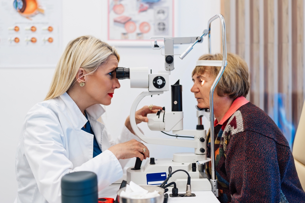
A comprehensive glaucoma eye exam looks at several distinct factors. No single test tells the whole story, which is why eye doctors typically use a combination of the following tests:
1. Tonometry
Tonometry measures the pressure inside the eye, known as intraocular pressure (IOP). Numbing drops are applied first, and then a small device gently touches the eye’s surface to take a reading. The average normal range is 10 to 21 mm Hg, though glaucoma can still develop with a normal IOP in some patients.
2. Optic Nerve Digital Photography
Optic nerve digital photography captures high-resolution images of the optic nerve. These photographs are compared over time, giving your eye doctor a visual record of the nerve’s appearance. Even subtle structural changes that might be difficult to detect in a single exam become apparent when images are reviewed side by side across multiple visits.
3. Optical Coherence Tomography (OCT)
Metro Eye Care also performs Optical Coherence Tomography (OCT) to measure how infrared light reflects off different tissues, producing precise images of the optic nerve fiber layer that can detect thinning before symptoms appear.
4. Perimetry
Perimetry, or visual field testing, maps a patient’s peripheral vision. During the test, lights of varying intensity appear in different parts of the visual field while the patient focuses on a central point. Any spots where perception is reduced are recorded and compared against age-specific norms. The procedure takes about 10 to 15 minutes and is repeated periodically to track changes over time.
5. Gonioscopy
Gonioscopy examines the drainage angle where the iris meets the cornea. This angle controls how fluid exits the eye, and when it’s blocked or too narrow, pressure can build. A hand-held lens is gently placed on the numbed eye to give the doctor a direct view of the angle’s structure.
6. Pachymetry
Pachymetry measures corneal thickness using a small probe placed briefly on the eye’s surface. This matters because corneal thickness can affect the accuracy of tonometry readings. A thinner-than-average cornea may cause the tonometer to underestimate actual pressure, while a thicker cornea may produce a higher reading than the eye’s true pressure.
Protect Your Vision with Metro Eye Care
Glaucoma screenings are quick, painless, and among the most effective tools available for protecting long-term vision health. The earlier the disease is detected, the more options there are to slow its progression.
If it’s been a while since your last comprehensive eye exam, or if you have risk factors for glaucoma, schedule a glaucoma screening at Metro Eye Care in Paramus, NJ, today!